3D digital cadavers Faculty Spark - View, reflect and apply
Last updated on 26/05/2020
-
You must be signed in to access this function
1
Description
Dr Leanne Kenway discusses how 3D models supplement student access to anatomy lab facilities in the School of Medical Science at Griffith University.
Challenge
We have an A-class anatomy facility and body donation program at Griffith University, and exposure to human cadaveric material is an essential component for students enrolled in my course.
One of the difficulties faced when teaching anatomy to our 1st year students is competition for space in our laboratory. With the increase in the number of courses utilising the facility, students really don’t have the opportunity to access the lab space for revision beyond their allocated lab session.
While the new Anatomy Learning Centre provides an excellent resource of models and texts for revision, students do not have access to the cadaveric specimens outside class time.
The nervous system is a module that is particularly content-heavy, and one that many students find more testing.
Approach
What I want to provide students with, is a supplement to their laboratory learning, accessible at any time, where they can revise content associated with the same cadaveric specimens that they see in the laboratory session, and that they will see again in their laboratory examinations.
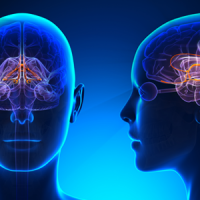
To address this, we’ve taken high-resolution 2-D images of elements of the nervous system, the brain and spinal cord, and through the use of software to process these images, we’ve produced 3-D rotating brains, and an online learning tool that contains questions associated with the 2-D images of these brains. These have been embedded within the 1016 MSC course site on Learning at Griffith, so that students may access these at any time, day or night, and make multiple attempts at these quizzes for revision purposes.
Outcomes
This blended learning strategy, incorporating 3-D digital brains as a supplementary learning tool aims to increase student engagement and improve performance outcomes in first-year anatomy courses.
Next Steps
The use of these 3-D digital brains will be evaluated through student satisfaction surveys, and by measuring performance outcomes in their laboratory exams. The level of student engagement will also be tracked through course analytics.
We plan to utilise this tool in more advanced anatomy courses to help scaffold knowledge, and for revision prior to commencing subsequent higher-level anatomy courses.
Support Resources
Contributed by
-
Learning Futures
-
Griffith Health
School of Medicine
Dr Leanne Kenway
(07) 555 29017
l.kenway@griffith.edu.au
http://orcid.org/0000-0001-7378-6612
Licence
© 2024 Griffith University.
The Griffith material on this web page is licensed under a Creative Commons Attribution NonCommercial International License (CC BY-NC 4.0). This licence does not extend to any underlying software, nor any non-Griffith images used under permission or commercial licence (as indicated). Materials linked to from this web page are subject to separate copyright conditions.
Preferred Citation
(2020). 3D digital cadavers. Retrieved from https://app.secure.griffith.edu.au/exlnt/entry/7448/view

